Orbital Trauma
Jump To
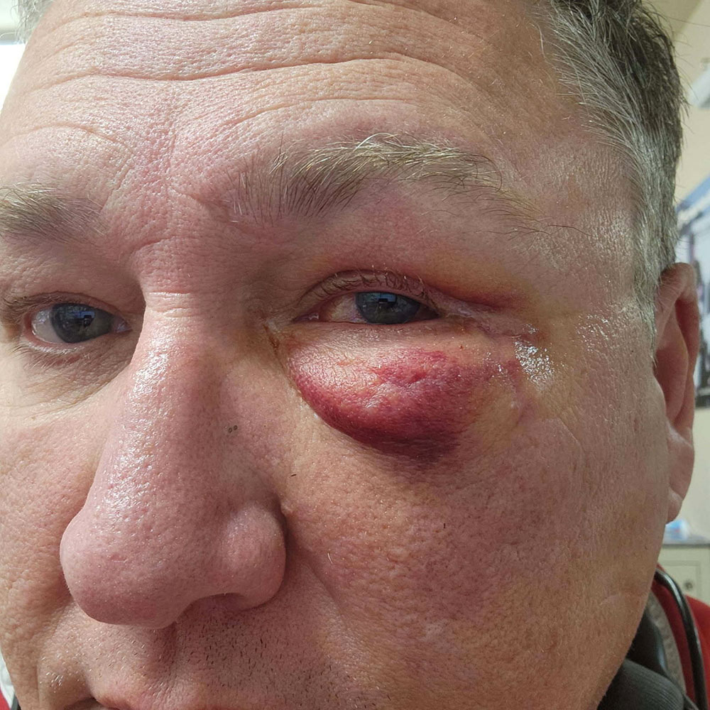
Ocular trauma can occur due to any injury impacting the eye and the area surrounding it (eyelid, eyeball, and supporting structures). Trauma can be caused by blunt force, accidents, punctures, foreign bodies, and chemical burns. Most often, symptoms include redness, swelling (edema), and pain, which can be accompanied by vision problems (blurred, double, or loss of vision). At Idaho Eyelid and Facial Plastic Surgery, our physicians diagnose ocular trauma by performing a visual acuity test, sometimes using CTs and MRIs, and by sometimes by examining the back of the eye. Treatment usually involves pain relief, antibiotics or anti-inflammatory drugs, and sometimes surgery to repair lacerations, remove foreign bodies, and restore vision.
Mid-facial orbital fractures involve the following: edema and ecchymosis of the soft tissues, subconjunctival hemorrhage, diplopia, iritis, retinal edema, ptosis, enophthalmos, ocular muscle paresis, mechanical restriction of ocular movement, and nasolacrimal disturbances. More severe injuries such as optic nerve trauma and retinal detachments can also occur. Intraocular injuries can occur when orbital bones are fractured.
Mechanism
Rates of injury have been compared between pure orbital floor fractures (only the floor) and floor/rim fractures (impure). Patients with pure orbital floor fractures had intraocular injuries at 5.6%, compared with only 2% in those who sustained an impure fracture. Intraocular injuries are more common in patients who sustained pure orbital fractures than in those with floor/rim fractures. This difference shows that the mechanism of injury is unlikely to be the same, as it could not simply be direct force transmitted to the rim, as the buckling theory suggests. Regarding intraocular injuries:
- The retropulsion theory refers to a fracture of the orbital floor caused by sudden increase in intra-orbital pressure, or a blowout. This happens when an object with sufficient force hits the aperture of the orbit and forces soft orbital tissue backward. This type of trauma generally occurs with objects larger than the horizontal diameter of the orbit. Examples include a fist or a ball.
- If impure fractures lead to direct trauma to the orbital rim and buckling of the floor, we might expect similar rates of intraocular injury between pure and impure orbital fractures. This theory stands if the buckling theory is correct for pure fractures, since both are created by the same direct force on the rim.
- The injury occurrence is much higher in the pure fracture group than in the impure group. This could point to a different mechanism. The acute rise in orbital pressure could lead to both floor fracturing and more intraocular injury. There is speculation, that with impure fractures, the rate of injury is lower because the force is blunted by the rim.
Buckling:
Buckling occurs when the orbital rim folds due to trauma and transmits forces to the orbital walls. This results in an orbital floor fracture.
Retropulsion:
The retropulsion concept was proposed by Smith and Regan. It suggests that orbital floor fractures are caused by sudden increase in intra-orbital pressure. Blowout from a fist, for example, or objects larger than the horizontal diameter of the orbit, are typical causes of this type of fracture.
Globe to Wall:
Globe to wall orbital fractures occur when the eyeball (globe) has been damaged to the point that its cornea and sclera (wall) have been punctured or penetrated. This can result in a full-thickness defect and is usually a medical emergency. This condition is known as either a globe rupture or open globe injury.
Consequences of trauma
Double Vision
Double vision can result from ocular trauma. It occurs when both eyes are unable to move equally because an anterior force is transmitted back into the orbit (buckling).
Surgery Indications
Ocular surgery may involve repairing or, in severe cases removing the eye, and sometimes treating damage to the cornea, lens, and retina. Globe repairs are typically performed within 24 hours of injury to close wounds, manage prolapsed tissue, and remove blood and foreign material. If there is additional damage, a camera-equipped probe can be utilized to treat media opacities and view the posterior segment of the eye. Surgery may be necessary to repair fractures that cause vision problems such as vision loss, double vision, or enophthalmos. Large (50%) floor fractures are likely to cause enophthalmos. Surgical repair is easier within the first two weeks from the trauma date. Surgical steps can include the following: inferior fornix incision, raising the periorbita from orbital floor, freeing tissues from fracture, and implanting (silastic or miniplate depending on size) over floor defect.
Medial Orbital Fracture
Surgery is not indicated if there is indirect (blowout) extension of the floor fracture. An exception occurs when the medial rectus is entrapped. Patients with direct naso-orbital fractures present a more serious situation. Under these conditions, patients may have a depressed nasal bridge, cerebral/ocular damage, severe epistaxis, and/or traumatic telecanthus.
Zygomatic Fracture
Zygomatic fractures relate to the cheekbone. Surgery involves a procedure to reduce and fix the cheekbone. It is usually recommended within three weeks of the injury. These injuries can involve the orbital floor and can result in cosmetic deformity.
Orbital Apex Fracture
The orbital apex is the posterior part of the orbit positioned at the craniofacial junction, located where the four orbital walls converge. Depending on the location and severity of the fracture, decompression may be needed. Some fractures don’t have to be treated immediately.
Orbital Roof Fracture
Orbital roof fractures occur when there is a break in the bone that forms the top of the eye socket. While rare, patients can have intracranial lesions, CSF rhinorrhea, or pneumocephalus. Sometimes, a neurosurgery consult is required.
Orbital Emphysema
Orbital emphysema occurs when a patient has air trapped in the tissue surrounding the eye orbit. This condition usually involves smaller medial wall injuries. If vision is decreased, high dose steroids are indicated, and there also may be a need to perform air decompression.
Treatment
While most fractures do not require surgery, patients should be evaluated after experiencing ocular or orbital trauma. Patients are often observed for a couple of weeks and given oral steroids (prednisone, for example) to decrease swelling and fibrosis. Antibiotics and nasal decongestants can also help. However, patients should not blow their noses to reduce the risk of orbital emphysema.